Red Florescent Protein Lab
Purpose: The main purpose of this lab was to learn the steps of genetic engineering; we used the same process of isolating a specific protein as major biotech companies would to make narcotics. Another purpose was to make red florescent protein from jelly fish in bacteria.
Materials: Micro-pipet(2-20 micro liters, 2-200 micro liters, 2-200 micro liters), Pipet tips, Micro-centrifuge tubes(1.5 mL), Sharpie, Mini centrifuge, Distilled water, Plasmid(pk), Reaction buffer(2.5x), Water bath, Ice container, Loading dye, R-, R+, SB buffer(50 mL of 1x), Agarose gel, DNA ladder, Gel box, Petri-dishes, Ampicillin, ARA-c, RFP, LB, Restriction enzyme, Gel tank, Luria broth(LB), E-coli cells(EC), Cell spreaders, Tape, Bio-hazard bag, Lysis buffer, Hydrophilic beads, Chromotography columns
Reason for Chemicals: RFP stands for red florescent protein and is the desired trait of the bacteria. Amp is an anti-biotic that is resistant to ampicillin. It shows the selection of bacteria that picks up the plasmid because only the bacteria with the recombinant plasmid grow on the Amp-agar. Without killing the nonresistant bacteria would be no visible way to differentiate the two pieces of bacteria because they would colonize together. ARA-c expresses the RFP protein that grows on the bacteria if it is placed on agar containing ARA. This is possible because ARA-c binds to the promoter of red florescent protein and forms messenger DNA from the DNA to produce the specific protein with the desired trait. The restriction enzyme cuts out a specific portion of the bacteria that contains the DNA of the trait. R- is the baseline control of a normal bacteria cell and R+ contains the bacteria with the restriction enzyme. This means R+ should have two cut pieces of the bacteria. LB stands for lysogeny broth and serves the purpose of providing a proper medium to support bacteria growth. This substance is spread onto specific perti dishes to control the results of an experiment.
Reason for Buffers: SB stands for sodium borate and therefore has low conductivity. This contrasts to TAE buffer because SB generates less heat and creates a lower temperature for the gels when an electric current is run through. This buffer was used in the verification of the recombinant plasmid. WB represents wash buffer. The purpose of using this is to wash the RFP proteins. EB is the elution buffer that knocks the protein off of the column in order to gather the desired protein as it runs out of the tube. CEB is the equilibration buffer that prepares the chromatography column for application. BB is the binding buffer that binds the proteins in the E-coli. These buffers were all used in the purification of the red florescent protein through chromatography columns.
Preparing to Verify the RFP-pARA-R Plasmid
Monday, November 3, 2014- Friday, November 7, 2014
Pre-Lab Questions: If the restriction enzyme is added to the sample of bacteria, the bacteria will be cut into two separate fragments with the size of eight hundred seven base pairs and four thousand four hundred ninety-five base pairs. The smaller fragment contains RFP and pBAD; the bigger fragment holds ARA-c, AmpR, and origins of replication. There are two components needed to create a plasmid that produces red florescent protein, RFP and ARA-c. The plasmid needs the actual red florescent protein and then the ARA-c expresses the protein. A selective marker is a gene placed in a cell to provide a specific trait that differentiates the cell from others. In this case, we used Amp to create bacteria plasmids resistant to ampicillin; only the bacteria that picked up the plasmid containing the RFP would also have the Amp. We can segregate the types of bacteria by placing all of them on a petri dish containing ampicillin so the bacteria with the plasmid won't colonize with the bacteria that does not contain the plasmid. This selective marker gives the bacteria a visible trait used to distinguish cells.
Procedure Part One: The exact procedure and materials list for this section can be found in Amgen Lab Manuel part 2a.
In this lab, BamH I and Hind III were used as the restriction enzymes that created the origin of replication. This is essential because it cut the RFP-ARA from the bacterial plasmid. Type-O.
In this lab, BamH I and Hind III were used as the restriction enzymes that created the origin of replication. This is essential because it cut the RFP-ARA from the bacterial plasmid. Type-O.
Post-Lab Questions: In order to clone a gene, you need the gene of interest, origin of replication, expression of gene with something that binds to the promoter, and a selective marker. A selective marker is a gene inserted into the bacteria to protect the organisms containing the plasmid from a specific agent in order to prevent cross colonizing. Restriction enzymes function as a defense mechanism in nature by destroying other harmful bacteria and foreign invaders. Bacteria retain genes with resistance to anti-biotics in order to survive and this affects the study of bio medicine. It requires us to find new advancements to fight against resistant bacteria; otherwise, humans would be susceptible to all bacterial diseases. Human genes can be expressed in bacteria even if the product has never been made in a bacteria cell because both prokaryotic and eukaryotic cells contain ribosomes. Ribosomes are essential because the organelle produces proteins from mRNA to express the trait. RNA polymerases are used to make the messenger RNA from the DNA in the organism. To separate bacteria with Amp and bacteria with kanamycin(anti-biotic) set four different petri dishes. The controlled variables will be the containers with both ampicillin and kanamycin and one with neither bacteria. Another dish will have only ampicillin so only bacteria with the Amp will grow on this plate; the final dish will hold kanamycin so only bacteria with the kanamycin resistant plasmid will grow. The expected growth on the plate containing both bacteria is for no colonies to form; the plate with neither of the bacteria should allow all of the bacteria to grow and should have the most number of colonies.
Verifying the Recombinant Plasmid Using Gel Electrophoresis
Monday, November 3, 2014- Friday, November 7, 2014
Pre-Lab Questions: Prior to the addition of the restriction enzyme, the bacteria should remain as circular plasmid. The configurations of the R- should be supercoil or circular plasmid because the bacteria should remain uncut due to the lack of restriction enzyme. The R+ should be cut plasmids that are smaller fragments. The supercoiled bacteria would have the most base pairs with over 5,300 bp, closely followed by circular plasmid that has approximately 5,000 bp. The fragment of ARA-c, ampR, and ori should have slightly less than 5,000 base pairs. Finally, the RFP and pBAD fragment should be the smallest with just over 800 bp. The largest particles with the most base pairs will travel the slowest through the gel. The RFP and pBAD fragment should be the closest to the bottom of the gel because it is the smallest plasmid and moves through the pores in the gel the fastest.
Procedure: The exact procedure and materials list for this section can be found in Amgen Lab Manuel part 4a.
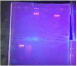
In order to verify the recombinant plasmid, we used gel electrophoresis to compare the size of fragments. A DNA ladder was loaded into one of the wells to display the size of specific fragments set at exact increments. This is used to find the size of the R- and R+ fragments. Unfortunately, our DNA ladder did not show, but we were still able to conclude that the R- fragments were larger than the R+ pieces.
In order to verify the recombinant plasmid, we used gel electrophoresis to compare the size of fragments. A DNA ladder was loaded into one of the wells to display the size of specific fragments set at exact increments. This is used to find the size of the R- and R+ fragments. Unfortunately, our DNA ladder did not show, but we were still able to conclude that the R- fragments were larger than the R+ pieces.
|
Calculations to Prepare Gel: (C1)(V1) = (C2)(V2)
(20x)(V) = (500 mg)(1x); V1 = 25 mL of 20x SB; therefore, 475 mL of water |
0.8% Agarose for 50 mL of water
(0.08)(50 mL)= 0.4 grams of Agarose |
Transforming Bacteria With the pARA-R Plasmid
Monday, November 3, 2014- Friday, November 7, 2014
Pre-Lab Questions: The purpose of growing bacteria on a plate with ampicillin is to ensure only bacteria containing the specific pARA-R plasmid will continue to grow. The bacteria possessing the ampicillin resistant plasmid should not be broken down by the ampicillin and should also hold the desired trait. If cells containing the pARA-R plasmid are not given arabinose, the gene will not be expressed because the cell will not produce the messenger RNA needed in order to form the protein.
Table of Predicted Bacteria Growth
|
Control (P-) Recombinant Plasmid (P+) |
Luria Broth
-Everything grows -Everything grows |
Luria Broth, Antibiotic Ampicillin
-All bacteria dies -Bacteria with plasmid grows |
Luria Broth, Antibiotic Ampicillin, Arabinose
-All bacteria dies -Gene expression in surviving bacteria |
Procedure: The exact procedure and materials list for this section can be found in Amgen Lab Manuel part 5a.
To transform the bacteria with the plasmid, we spread bacteria onto several variations of petri dishes and observed the results. Different quantities of bacteria colonies developed and displayed the RFP gene depending on the type of plate and if the bacteria held the plasmid.
To transform the bacteria with the plasmid, we spread bacteria onto several variations of petri dishes and observed the results. Different quantities of bacteria colonies developed and displayed the RFP gene depending on the type of plate and if the bacteria held the plasmid.
Post-Lab Questions: For the majority of the plates, the expected amount of bacteria grew. Unfortunately, on the LB/Amp/ARA petri dish, the red florescent protein was not expressed despite the number of bacteria colonies that grew on the plate. This error was caused by the incorrect RFP gene being sent to our school because the company mixed up the order for materials and the change of results was not due to human error during the procedure. On the LB/Amp/ARA plate, there were no red colonies, but there were still non-florescent bacteria colonies growing. However, if red colonies did appear on this petri dish, it would be because it was the only arrangement that contained ARA, which is essential in creating mRNA that forms the protein to express the gene. It is important for multiple copies of the recombinant gene to be made within a cell so there are backup replications in case one recombinant plasmid dies. The RFP gene can be expressed as a trait because the central dogma. The ARA creates messenger RNA from the DNA in the RFP gene. This mRNA can form protein and the specific protein built displays the desired trait. Bacteria is able to create human proteins because bacteria and human cells both have similar components. Both types of cells posses ribosomes that produce the proteins needed to express the traits of the genes, regardless if it is a human protein.
Purification of Red Florescent Protein
Monday, December 8, 2014- Wednesday, December 17, 2014
Stop and Think Questions: You are able to determine the position of the red florescent protein by observing the color of the e-coli or beads. The pink or red coloration is a characteristic that indicates the protein. The supernatant was clear and was composed of liquid with the nutrients such as amino acids and sugars that cells need to grow. The pellet was pink because it had the RFP cells. The function of each buffer we used is located at the top of the page under the section titled Reasons for Buffers. After running the buffers through the chromatography column, the supernatant was light pink and held e-coli. The pellet was a dark pink that looked almost red and contained RFP cells.
Procedure: The exact procedure and materials list for this section can be found in Amgen Lab Manuel part 6a.
We used chromatography columns and several different buffers to purify the RFP. Throughout this section of the lab, we recorded the color of the supernatant and pellet in order to track the placement of the red florescent protein cells. The chromatography columns that we used included hydrophilic beads to attract the molecules in the RFP.
We used chromatography columns and several different buffers to purify the RFP. Throughout this section of the lab, we recorded the color of the supernatant and pellet in order to track the placement of the red florescent protein cells. The chromatography columns that we used included hydrophilic beads to attract the molecules in the RFP.
To people in the future, I did plan on uploading a picture of our verification gel along with notes concerning the results, but all my pictures on my phone got deleted when I tired to sync it to my computer, so there will not be any extra photos until I am able to recover the data.
